DIAGNOSIS AND TREATMENT
Argon Laser: Ruptures in retina layer, holes, bleeding due to diabetes, abnormal blood vessels and vessels that lead to leakage can be treated with argon laser. Also argon laser can be used in intraocular pressure.
Yag Laser: It is the laser type used in the opening of the posterior capsule that is disintegrating after cataract surgery and in the treatment of some glaucoma types.
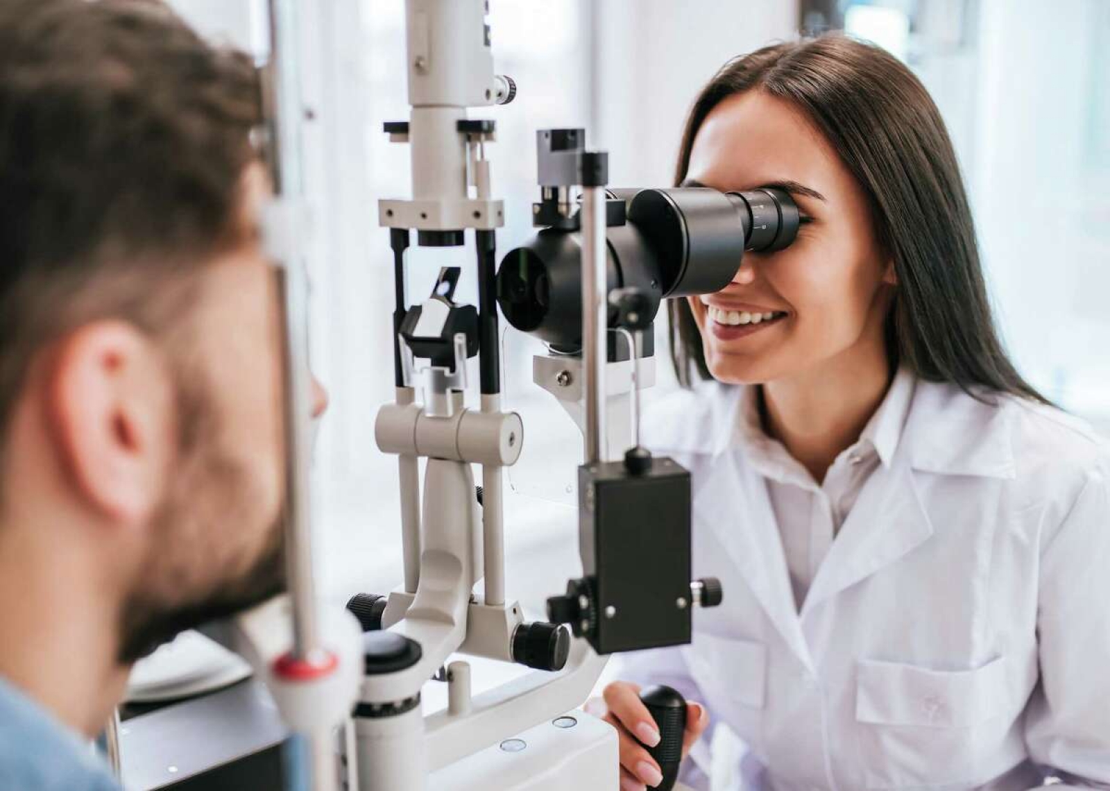
Ultrasonography (USG): It is used to visualize posterior segment of the eye in conditions that are not visible with normal examination devices during normal eye examinations.
Ocular Biometry: It is used for eyeball measurements and to determine intraocular lense degree.
Optical Coherence Tomography (OCT): Used in diabetes, macular degeneration disease and retinal diseases. This is the only way that shows the thickness of nerve fibers in glaucoma disease.
Pachymetry: Measuring thickness of the transparent layer (cornea) in front of the eye.
Fundus Fluorescein Angiography (FFA): Used in the diagnosis and monitoring of retinal diseases by allowing the examination of retinal blood flow. Fundus Fluorescein Angiography is used diagnosis and differential diagnosis of many retinal diseases as well as diabetic retinopathy, retinal vascular occlusions, age-related macular degeneration, inflammatory retinal diseases.
Visual Field Analyzer (VFA): It is used for examining the damage that eye tension and some brain lesions may cause in the visual nerve.
Contact Lens: Lenses are attached to the front surface of the cornea as an alternative to eyeglasses to fix eye defects.
Plusoptix: Makes early detection possible by measuring eye defect of babies.
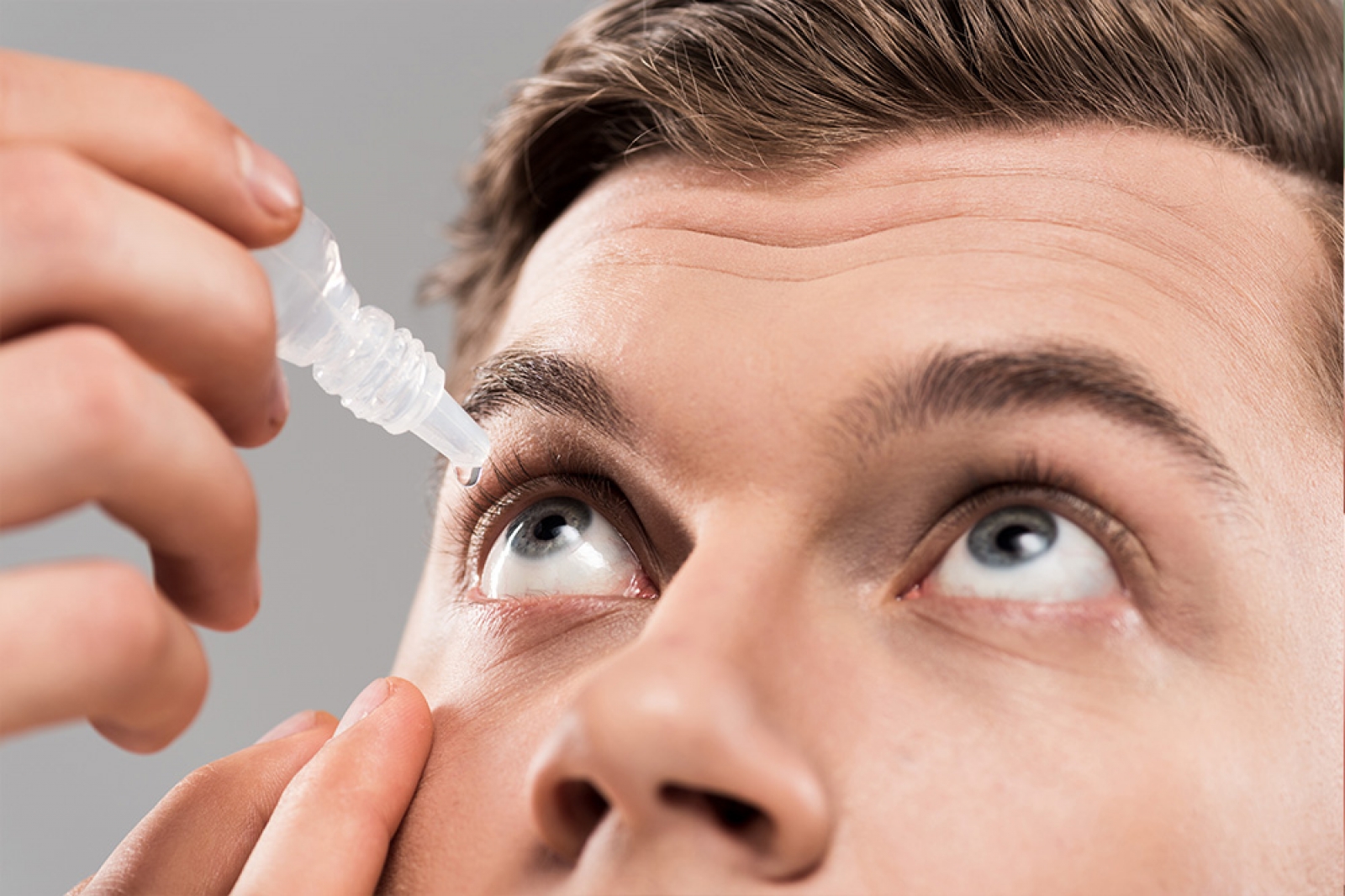
Intraocular drug injections: Injections into the vitreous to provide the drug accessing directly in the eye. Intraocular drug injections can be applied in the treatment of severe intraocular infections such as age related macular degeneration, diabetes mellitus, water collection in yellow spot due to retinal vascular occlusion (macula edema) and endophthalmitis.
EYE SURGERIES
Eye surrounding lesions, Strabismus, Glaucoma, Cataract (FACO) + intraocular lens operations are performed.


 info@osmanogluhastanesi.com.tr
 +90 850 800 50 50
 WhatsApp


